| Home News NeuroMabs Protocols Publications BRAIN Initiative Contact Us | |
|
|
| Welcome to NeuroMab! |
|
The UC Davis/NIH NeuroMab Facility is a purely academic monoclonal antibody (mAb) facility with a mission to generate mouse mAbs extensively validated for use in mammalian brain, as funded by NIH awards U24 NS050606 (2005-2015), R24 NS092991 (2015-2019) and U24 NS109113 (2019-present). The NeuroMab collection (both the mAbs and the hybridoma cells that produce them) is available from the Developmental Studies Hybridoma Bank (DSHB) at the University of Iowa. NeuroMab hybridomas are also available from the UC Davis MMRRC. Recombinant mAbs derived from the NeuroMab collection are available from Addgene as plasmids and, as funded by BRAIN Initiative award NIH U24 NS119916, as ready-to-use antibodies. The sequences of the heavy and light chain variable domains of most NeuroMabs are now available on the NeuroMabSeq website here. These sequences were obtained by high-throughput sequencing of hybridoma-derived RNA as funded by BRAIN Initiative award NIH U24 NS109113. See our publications here and here. NeuroMabs are validated for biochemical and immunohistochemical applications in mammalian brain samples, including immunoblotting, immunohistochemistry and array tomography. For information on NeuroMab validation see Gong et al., New Biotech 33:551, 2016. PubMed. See detailed sample preparation and labeling protocols used for NeuroMab monoclonal antibody validation. NeuroMab is a proud supporter of the Research Resource Identification (RRID) initiative. Our unique identifier is RRID:SCR_003086. For perspectives on application-specific antibody validation see Taussig et al., New Biotech 45:1, 2018. PubMed. See publications list for peer-reviewed papers showing data obtained with NeuroMabs, now over >6,300 publications! 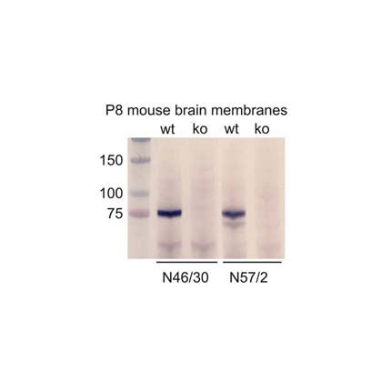 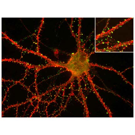 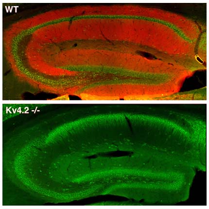 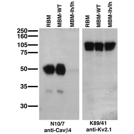 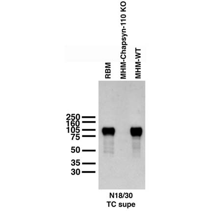 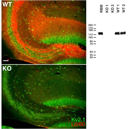 Immunoblot against membranes from postnatal day 8 wild-type (wt) and ADAM22-knockout (ko) mice probed with N46/30 (left, ADAM22-cytoplasmic) and N57/2 (right, ADAM22-extracellular). Data courtesy of Dies Meijer (Erasmus University Medical Center). Immunofluorescence staining of cultured rat hippocampal neurons with K28/43 (green, PSD-95) and K57/1 (red, Kv4.2) Immunofluorescence staining of adult wild-type (WT, top) and Kv4.2 knockout (Kv4.2-/-, bottom) mouse hippocampus with K57/1 (red, Kv4.2) and Kv2.1 rabbit polyclonal (green) Immunoblot against brain membranes from adult rat (RBM) and adult wild-type (MBM-WT) and lethargic (MBM-lh/lh) mice probed with N10/7 (left, Cavbeta4) and K89/41 (right, Kv2.1). Lethargic mice courtesy of Jeffrey Noebels (Baylor College of Medicine). Immunoblot against membranes from adult rat brain (RBM) and hippocampi from adult wild-type (MHM-WT) and Chapsyn-110 knockout (MHM-Chapsyin-110 KO) mice probed with N18/30 (Chapsyn-110). Mouse samples courtesy of Richard Huganir (Johns Hopkins University, Howard Hughes Medical Institute) Immunofluorescence staining of hippocampal CA3 of adult wild-type (WT, left top) and Slo1 knockout (KO, left bottom) mice with L6/60 (red, Slo1) and Kv2.1 rabbit polyclonal (green). Immunoblot against brain membranes from adult rat (RBM) and adult WT and Slo1 KO mice probed with L6/60 (right, Slo1) |